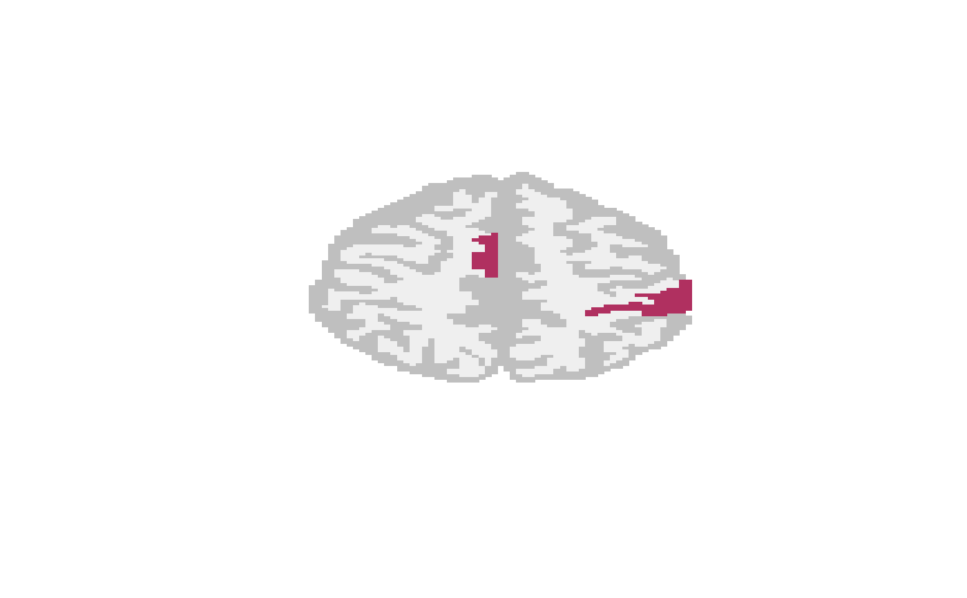
A \(128 \times 128\) activation map for a slice of an fMRI phantom and an anatomical reference.
data(hoffmanphantom)Format
a \(128 \times 128 \times 2\) array with the
first slice an activation map for an MRI phantom and the second an
anatomical overlay. NA values are outside the surface. The
activation map (hoffmanphantom[,,1]) is 1 if activated,
0 otherwise. The second layer (hoffmanphantom[,,2])
indicates the anatomical structure. Approximately 3.8 percent of the
pixels are activated in this slice.
References
E. Hoffman, P. Cutler, W. Digby, and J. Mazziotta, “3-D phantom to simulate cerebral blood flow and metabolic images for PET,” Nuclear Science, IEEE Transactions on, vol. 37, pp. 616 – 620, 05 1990.
I. A. Almodóvar-Rivera and R. Maitra, “FAST adaptive smoothed thresholding for improved activation detection in low-signal fMRI,” IEEE Transactions on Medical Imaging, vol. 38, no. 12, pp. 2821–2828, 2019.